Hyperplastic polyp of stomach with intestinal metaplasia


Juvenile rectal polyp
 |
| Cribriform appearance on low power |


More, from lower magnification


Colonic adenoma

 |
| Dysplasia is a requisite criterion |
|
| Cribriform appearance on low power |
|
| Dysplasia is a requisite criterion |


 |
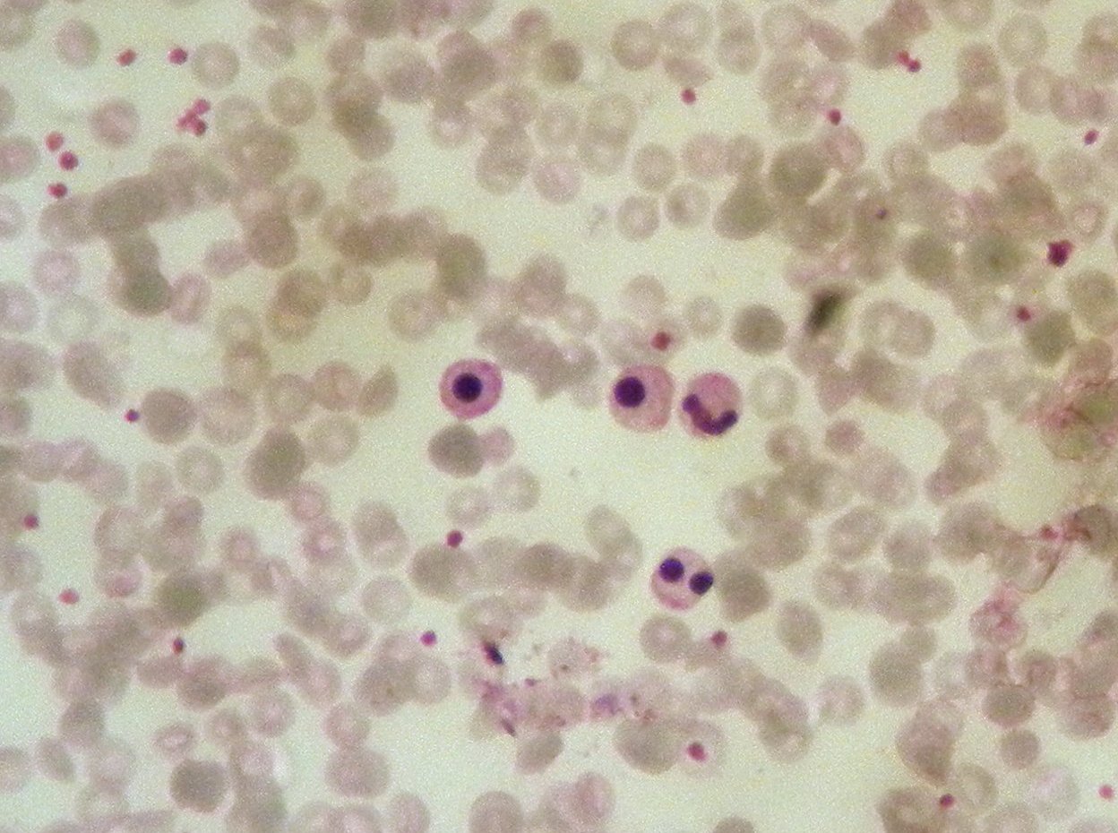
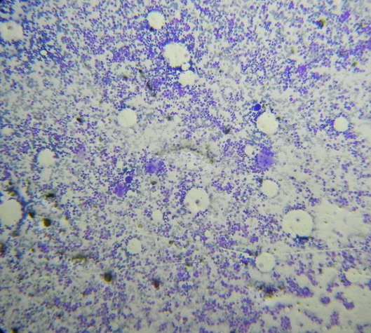
| Note the tapering WBC histogram |
 |
| PBS |

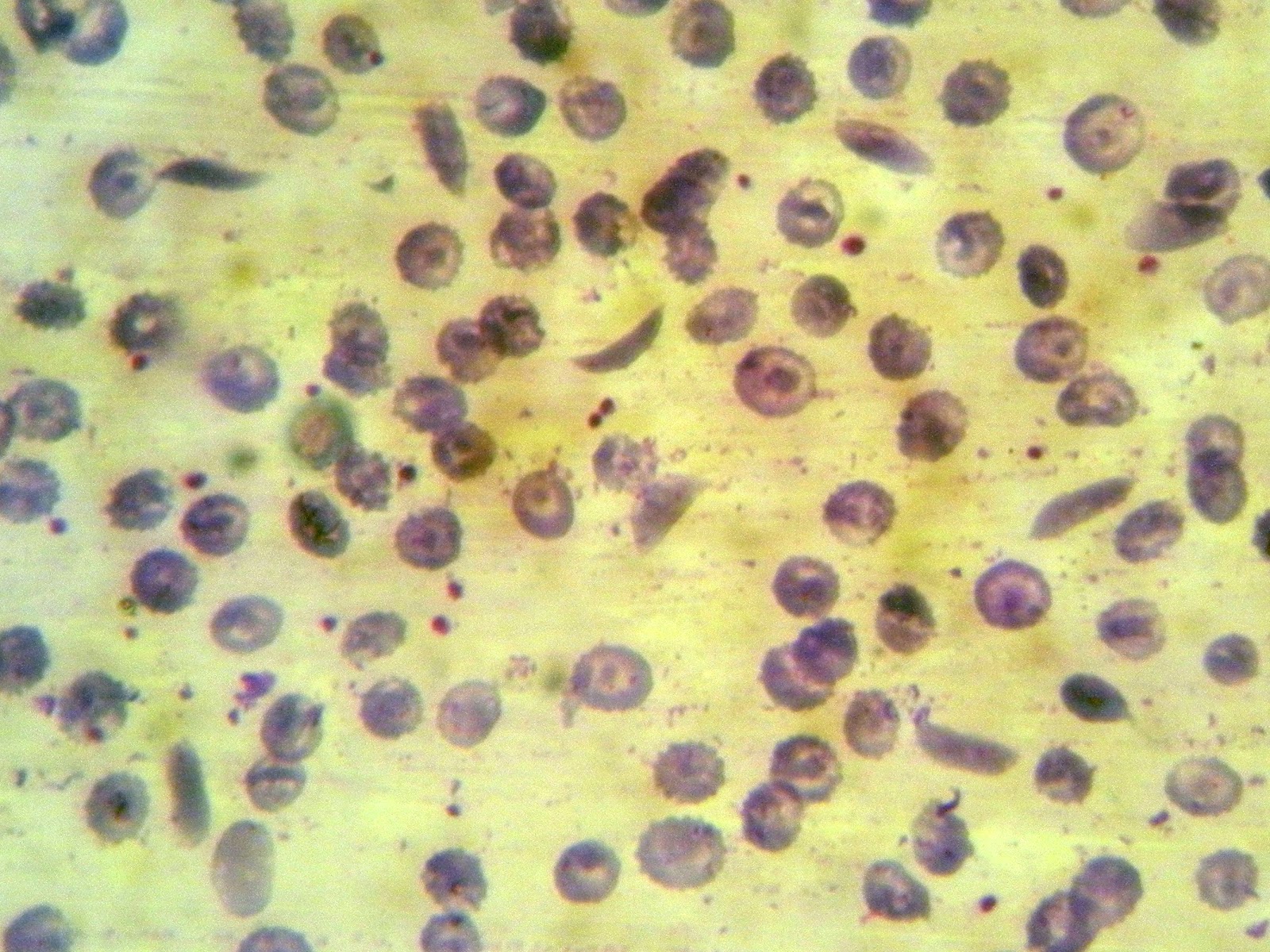 |
| PBS |


 |
| The dark staining exocrine pancreatic lobules surrounding an islets of langerhans. A duct is seen in right. |
 |
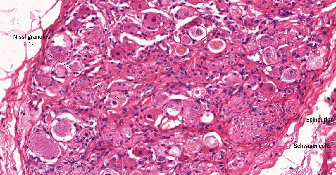
| Islet of Langerhans |
 |
| Cut surface is a blackish cyst |
 |
| Showing normal pancreas (left) with neoplasm in right |
 |
| Solid pseudopapilary neoplasm |
 |
| Blackened pancreas |
 |
| Blackened omentum around pancreas |
 |
| Fat necrosis around pancreas |
 |
| Left - pancreas; right - saponified fat |


 |
| Saponification |
Imagine solving a puzzle with 100 pieces, each piece a centimeter in size, something like this: The genome is considerably larger than this...


