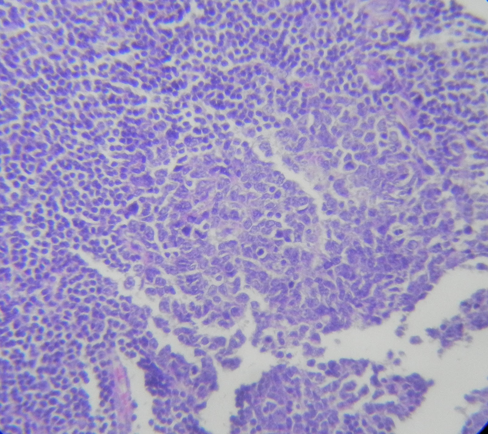
A lymph node. Disrupted architecture? Some extracapsular invasion, maybe?




 |
| Atrophy? LSIL? More like the former. |
 |
| The jigsaw in the dermis. A cylindroma from scalp. |
 |
| The customary storage disease slide from liver |
 |
| Looks like crumpled tissue |
 |
| Bone marrow with lot of macrophages |
 |
| which are devouring RBCs. Hemophagocytic lymphohistiocytosis. |
 |
| Skin |
 |
| Strain your eyes to see the pink granules in between cells. Round fungi, possibly histoplasma. |
 |
| The proceedings |
 |
| Hemolysis. History suggestive of microangiopathic hemolytic anemia. |

 |
| Mixed cellularity Hodgkins |
 |
| Neuroendocrine neoplasm. Breast |
 |
| A thyroid well dermarcated thyroid lesion, with pigmented regions |

 |
| Pigmented medullary carcinoma (!) |
 |
| Plamsacytoid cells in PBS but with an open nucleus |
 |
| Plasmablastic leukemia? |
 |
| Minor salivary gland. Polymorphous low grade adenocarcinoma. |
 |
| Lymph node. |
 |
| Lot of macrophages. |
 |
| Emperipolesis (em = inside, peri=around, polemal = wander about) |
 |
| Rosai Dorfman disease |
 |
| Testis FNAC |
 |
| Tigroid background; large round cells with streaked out chromatin |
 |
| Classical seminoma |


