Gleason’s grading is done only for primary cancers (not metastatic deposits) and those who are unmodified by chemotherapy or radiotherapy. It is the only known architectural grading system, as opposed to other cytological ones. It is also unique that it is reported as three scores, primary, secondary (second common pattern) and total.
Originally, Donald Gleason described 9 patterns that have been stratified into 5. However, the latest IUSP grading abolishes the subgrades and keeps only 5 (no more 4a and 4b, just say '4') . Small cell cancer and paneth cell like adenocarcinoma are NOT graded.
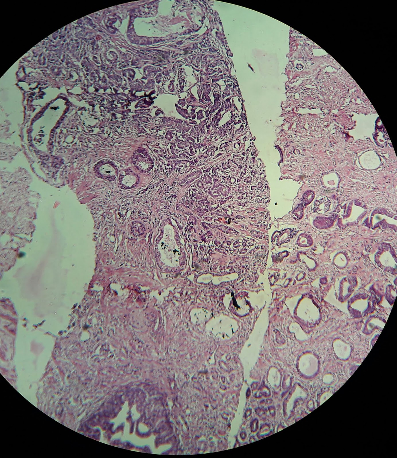
Intraepithelial neoplasia
Low grade PIN
Only nuclear hyperchromasia
High grade PIN
Nucleoli visible at 20X (> 1 μm). May show architectural features like cribriform/ tufted/ micropapillary.
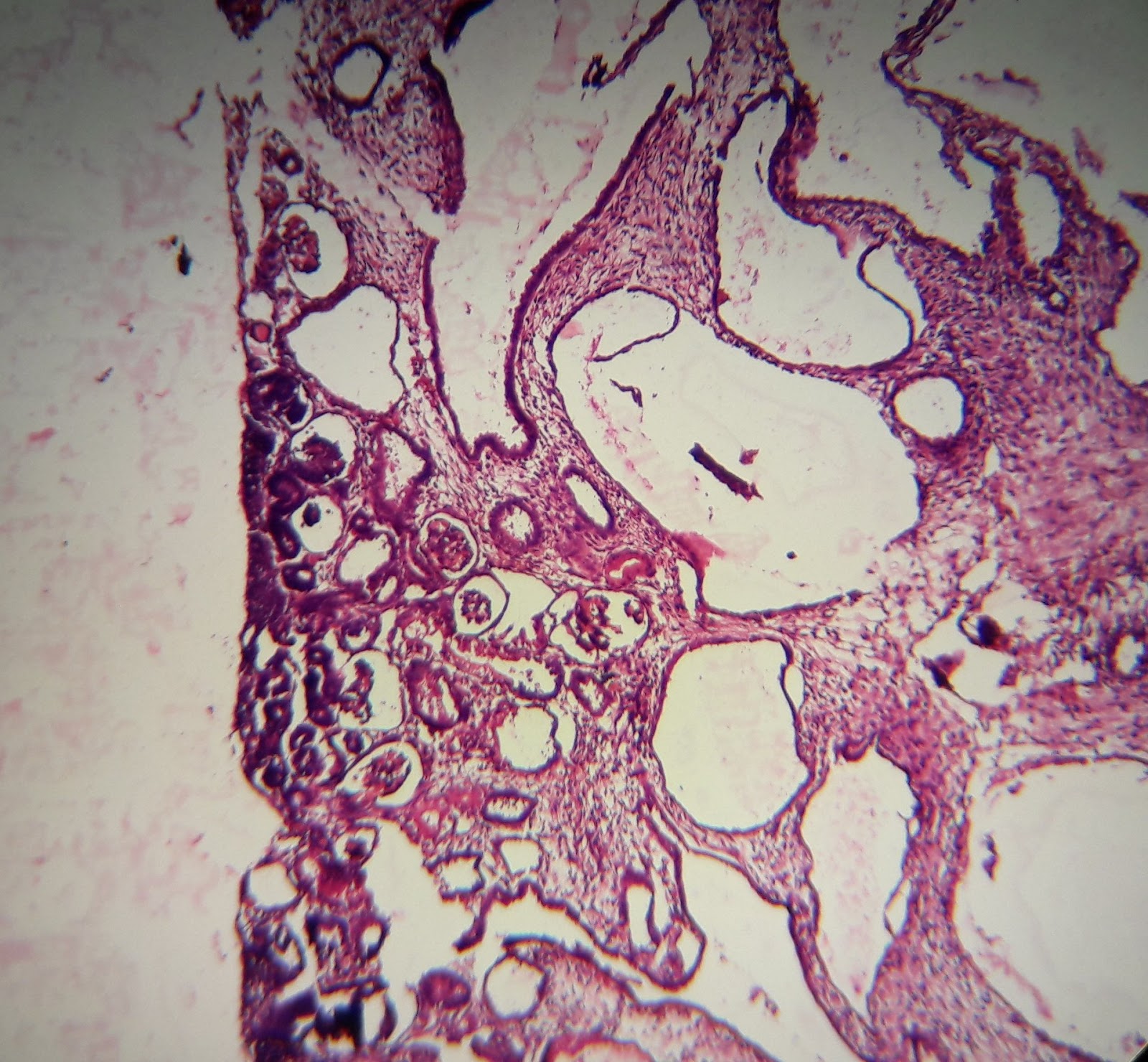
 |
| Intraepithelial neoplasia - cribriform |
Gleason 1
Round to oval, back to back glands in
well circumscribed nodules (no invasion allowed); rare. Should NEVER be reported on a core biopsy, because circumscription can rarely be made out in a core biopsy. Even on TURP or prostatectomy specimens, it is sufficiently rare.
Gleason 2
Well circumscribed nodule but glands are looser, more variable in size with minimal infiltration. The stroma is more than pattern 3, in this case more than one glandular space. This too is rare. In a needle biopsy, report otherwise circumscribed nodules as grade 2, because you never know where they could be infiltrative.
Gleason 3
Infiltrative pattern, anisoglandulosis, angular glands, but glands are still distinct; gland spacing < one gland diameter. Occasional V or Y shaped gland can mimic fused glands, but try drawing a line around each gland and you’ll find they are distinct. Glands might be medium, small with hardly any lumen (commonest pattern) or cribriform / papillary ('papillary intraductal tumour').
 |
| Gleason 3 |
 |
| Medium and small glands |
 |
Involving a nerve
|
This ones showing cribriform pattern in a small focus
 |
| On closer look, the cells are bad looking |
Gleason 4
Glands become fused, poorly defined, cribriform or glomeruloid, raggedly outlined, raggedly infiltrating fused glands. In some cases, the fused glands become clear giving a clear cell carcinoma like ('hypernephroid') apperance. Occasionally, this can be confused with foamy gland carcinoma
Gleason 4 + 3
 |
| Note fusion of glands |
Gleason 4 again
 |
| Completely fused glands, but can still be made out as glands |
Gleason 5
No glands, solid sheets of cells, rosettes, cords or single cells OR
comedonecrosis; cribriform pattern can also be seen. The cancer may be so poorly defined as having no glands but only single cells and strands (can be mimicked by small cell carcinoma)
The sum of dominant and subdominant pattern is given as combined Gleason score.
Gleason 5 + 4
 |
| Mainly single cells in sheets |
Gleason 5 again
 |
| Solid clusters of cells |